Joint Preservation Surgery Using Arthroscopy
Joint Preservation Surgery Using Arthroscopy
Joint preservation through arthroscopy is a cutting-edge approach designed to treat early to moderate joint damage while maintaining your natural joint structure. Using a tiny camera and specialized instruments, orthopedic surgeons can visualize, diagnose, and treat internal joint problems with precision — all through small incisions.
Causes of Joint Damage
1. Traumatic Injuries
Accidents, falls, or sports injuries can result in fractures, dislocations, ligament tears, or cartilage damage — all of which can compromise joint stability and function.
2. Degenerative Conditions
Chronic diseases like osteoarthriti rheumatoid arthritis, or avascular necrosis cause the gradual breakdown of cartilage and bone, leading to joint deterioration.
3. Overuse Injuries
Repetitive stress from work, sports, or improper biomechanics can cause microtrauma that accumulates over time, wearing down cartilage and soft tissues.
4. Congenital Abnormalities
Structural problems present from birth — such as hip dysplasia or limb deformities — can lead to uneven joint stress and early degeneration.
Indications for Joint Preservation Surgery
1. Chronic Pain
Persistent or worsening joint pain that limits daily activities and does not respond to non-surgical interventions.
2. Joint Stiffness
Difficulty in moving the joint due to internal damage, scar tissue, or cartilage loss.
3. Swelling and Inflammation
Recurrent joint swelling that leads to discomfort and impaired function, often linked to synovial irritation or cartilage damage.
4. Decreased Range of Motion
Inability to fully bend, stretch, or rotate the joint — affecting sports, work, or basic movements like walking or reaching.
Diagnosis of Joint Damage
Identifying the extent and cause of joint damage is the first step toward effective treatment and joint preservation. A thorough diagnostic process ensures that the right surgical or non-surgical plan is selected for optimal outcomes.
1. Clinical Evaluation
An orthopedic specialist will begin with a detailed medical history and physical examination to assess:
Pain level and location
Joint mobility and stiffness
Swelling, instability, or deformity
Impact on daily activities and movement
2. Imaging Studies
Imaging tests help visualize internal joint structures and identify cartilage, bone, or soft tissue damage.
X-rays: Detect bone deformities, arthritis, fractures, and alignment issues
MRI (Magnetic Resonance Imaging): Provides high-resolution images of cartilage, ligaments, tendons, and menisci
CT Scan: Offers detailed cross-sectional views of bone and joint surfaces
Ultrasound: Useful for assessing soft tissue injuries and joint inflammation in real time
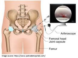
3. Diagnostic Arthroscopy
In certain cases, minimally invasive arthroscopy may be used not only for treatment but also to directly view the joint interior. It allows:
Precise identification of cartilage lesions
Assessment of ligament integrity
Confirmation of damage extent when imaging is inconclusive
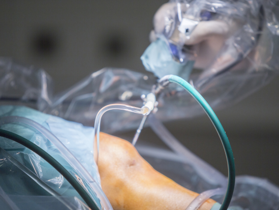
Arthroscopic Techniques for Joint Preservation Surgery
Arthroscopy is a keyhole surgical technique that allows orthopedic surgeons to diagnose and treat joint damage with high precision while minimizing trauma to surrounding tissues. It plays a vital role in joint preservation surgery, helping to delay or avoid the need for total joint replacement.
1. Chondroplasty
Smooths rough or damaged cartilage to reduce friction and inflammation.
1. Ideal for early-stage cartilage wear.
2. Microfracture Surgery
Small holes are drilled in the bone beneath damaged cartilage to stimulate new cartilage growth.
1. Effective for small, contained cartilage defects.
3. Autologous Chondrocyte Implantation (ACI) (Advanced)
Cartilage cells harvested from the patient are cultured in a lab and later re-implanted into the damaged area.
1. Used for larger defects in younger patients.
Benefits of Arthroscopic Joint Preservation Surgery
1. Minimally Invasive Approach
-
Small incisions mean less trauma to surrounding tissues.
-
Reduced scarring and improved cosmetic outcomes.
2. Faster Recovery Time
-
Most patients experience quicker rehabilitation compared to open surgery.
-
Shorter hospital stays or same-day discharge in many cases.
3. Less Postoperative Pain
-
Reduced tissue disruption results in lower pain levels after surgery.
-
Less reliance on pain medications.
Consult Us
We specialize in advanced arthroscopic joint preservation surgeries to help you maintain mobility, reduce pain, and avoid early joint replacement.
Whether you’re experiencing knee, shoulder, or hip discomfort — or recovering from a sports injury — our expert orthopedic team is here to guide you every step of the way.